- Visibility 33 Views
- Downloads 11 Downloads
- DOI 10.18231/j.ijmi.2021.029
-
CrossMark
- Citation
A cross-sectional analysis of digital panoramic radiographs for the evaluation of elongated styloid process in central Kerala population
- Author Details:
-
Raina Susan Reji *
-
Sheeba Padiyath
-
Anu Vijayan
-
Anju Elizabeth Thomas
-
Sonia Susan Philip
Introduction
The styloid process is a cylindrical, long cartilaginous bone located on the temporal bone. It arises from under surface of petrous part of temporal bone in front of stylomastoid for a men.[1] The normal length of styloid process is 20-30 mm, but it varies from person to person and side to side within the same person.[2] A styloid process is said to be elongated, if the overall length exceeds 30 mm.[3] The prevalence of an elongated styloid process in general population is about 2-28%.4Elongated styloid process is known as Eagles Syndrome when there are clinical manifestations such as neck and cervico facial pain. It is believed that these signs and symptoms arise due to compression of styloid process on some neural and vascular structures.[1] These symptoms can be confused with a wide variety of facial neuralgias, oral and temporomandibular disorders. Thus, it is important to have a precise knowledge about the anatomy of both normal and abnormal styloid process for clinicians, surgeons and radiologists. Panoramic radiography is a commonly used modality for evaluation of styloid process.
Materials and Methods
The aim of the study was to investigate the prevalence, morphology and elongation pattern of elongated styloid process in the population of Central Kerala and its relation to age and gender. A cross-sectional study was conducted on 500 digital panoramic radiographs collected from the archives of Department of Oral Medicine & Radiology, Mar Baselios Dental College taken during 2017-2020. Only those radiographs of patients within age group of 20-60 years and showed styloid processes of both sides with no pathologies in the required structures were included in the study. Radiographs with magnification and positioning errors were excluded.
The radiographs were taken in a digital panoramic system. The length of styloid process was measured as distance from the point where the styloid process left the tympanic plate to the tip of the process. Styloid process measuring more than 30 mm was considered to be elongated. The type of elongation of styloid process based on the pattern of calcification were classified as per Langlais.
The collected data was entered in an Excel spreadsheet and analyzed using statistical analysis software-SPSS 16.0.
Results
In the 500 panoramic radiographs studied, a total of 1000 styloid processes were evaluated.
|
Group Statistics |
|
|
|
|
|
|
|
|
|
Groups |
N |
Mean |
Std. Deviation |
t |
Sig. (2-tailed) |
Mean Difference |
|
Length |
Left |
500 |
25.6143 |
6.25593 |
|
|
|
|
|
Right |
500 |
25.8807 |
6.67751 |
-0.651 |
0.515 |
-0.26646 |
The mean length of styloid processes in left side is 25.6143±6.25593 mm and mean length of styloid processes in right side is 25.8807±6.67751 mm[Table 1]. The difference between length of styloid processes in left side and right side was not statistically significant[p-value 0.515>0.05]
|
Group Statistics |
Gender |
Mean |
Std. Deviation |
t |
df |
Sig. (2-tailed) |
Mean Difference |
|
|
Length of Left Styloid Process |
Male |
193 |
25.9693 |
5.88989 |
1.006 |
498 |
0.315 |
0.5782 |
|
Female |
307 |
25.3911 |
6.47493 |
[Table 2] Shows that the mean length of left styloid processes in males were 25.9693±5.88989 mm and mean length of left styloid processes in females were 25.3911±6.47493 mm. The difference between length of left styloid processes in males and females were not statistically significant[p-value 0.315>0.05]
|
|
|
|
|
|
|
|
|
|
|
Group Statistics |
Gender |
N |
Mean |
Std. Deviation |
t |
df |
Sig. (2-tailed) |
Mean Difference |
|
Lenght of Right Styloid Process |
Male |
193 |
26.9199 |
6.75934 |
2.778 |
498 |
0.006 |
1.69247 |
|
Female |
307 |
25.2274 |
6.55253 |
[Table 3] Shows that the meanlengths of right styloid processes in males were 26.9199±6.75934 mm and mean length of right styloid processes in females were 25.2274±6.55253 mm. The difference between lengths of right styloid processes in males and females were statistically significant [p-value-0.006<0.05]
Females had a longer styloid process compared to males.
|
Correlations |
|
|
|
|
|
|
Age |
Length |
|
Age |
Pearson Correlation |
1 |
.123** |
|
|
|
Sig. (2-tailed) |
0.006 |
|
Length |
Pearson Correlation |
.123** |
1 |
|
|
|
0.006 |
|
[Table 4] Shows that there was a positive correlation between age and length of left styloid process.
|
Correlations |
|
|
|
|
|
|
Age |
Length |
|
Age |
Pearson Correlation |
1 |
.218** |
|
|
|
Sig. (2-tailed) |
0.00 |
|
Length |
Pearson Correlation |
.218** |
1 |
|
|
|
0.00 |
|
[Table 5] Shows that there was a positive correlation between length of right styloid process and age.
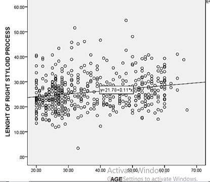
The average length of right styloid process showed an increasing pattern as the age increased.
|
Type of Elongation |
Frequency |
Percentage |
|
Type I |
409 |
81.8 |
|
Type II |
50 |
10 |
|
Type III |
23 |
4.6 |
|
Type IV |
18 |
3.6 |
|
Type of Elongation |
Frequency |
Percentage |
|
Type I |
389 |
77.8 |
|
Type II |
67 |
13.4 |
|
Type III |
22 |
4.4 |
|
Type IV |
22 |
4.4 |
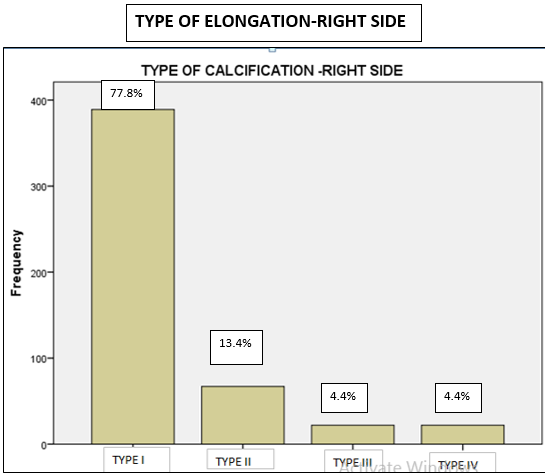
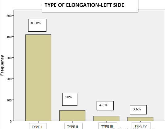
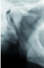
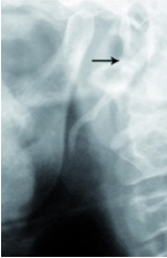
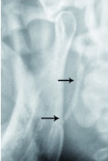
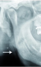
[Table 6], [Table 7] Shows that Langlais Type 1 elongation[[Figure 4]] had a predominance in both sides[81.8 % in left & 77.8% in right]. Type II elongation[[Figure 5]] was found to be 10% in left side & 13.4% in right side. Type III elongation [[Figure 6]] was about 4.6% in left side & 4.4% in right side. Type IV elongation[[Figure 7]] was about 3.6% in left side & 4.4% in right side.
Discussion
It is very important for all clinical practitioners to have an awareness about clinical and radiologic presentation of styloid process elongation as they are involved in the diagnosis and treatment of head and neck pain. Eagle syndrome or styloid syndrome or stylohyoid syndrome is the symptomatic elongation of the styloid process or mineralization {ossification or calcification}.[4] It was first put forward by Eagle, an otorhinolaryngologist in 1937.[2] The styloid process tapers towards its tip that lies in the pharyngeal wall lateral to the tonsillar fossa. If styloid process is palpable in the tonsillar fossa, which is not normally palpable is an indicative of styloid process elongation and it can be confirmed by radiologic imaging.
Panoramic radiograph is a common modality for evaluating styloid process elongation. Lateral skull view is the best to evaluate length of styloid process but anteroposterior views are also required to determine whether there is bilateral involvement and presence of lateral deviation. For exact determination of localization of styloid process, spiral CT with subsequent 3D reconstruction is the method of choice.[5] Apart from Eagle syndrome; other manifestations of elongated styloid processes are glossopharyngeal neuralgia, carotidynia, pulsatile tinnitus, and dysphonia & globus pharyngeus.[6]
In the present study, the average length of right and left styloid processes were 25.61±6.25mm & 25.88±6.67 mm respectively. Eagle [1948][7] reported that length of normal styloid process is app.2.5-3 cm whereas Kaufman et al. [1970][8] concluded 30mm as the upper limit for normal styloid process. Bozkir et al.[1999][9] studied panoramic radiographs of 200 edentulous patients above 50 years of age, and found average length to be 53mm.Thot et al.[2000][10] reported average styloid process length to be 1.49 cm on the right and 1.52 cm on the left. Jung et al.[2004][11] stated that for the styloid process to be considered as elongated, the length should exceed 45 millimeters. The differences in the results of our study may be due to differences in age groups and sample size.
The study showed that there is a progression in the length of right styloid process with advancing age. This finding was similar to the results of other studies reported in literature by More and Asrani [2010].Vajendra Joshi [2007] evaluated 62 panoramic radiographs in the age group of 10-70 years and reported a similar finding.[12] This was also in harmony with a review by Jaju PP[2007].[13] In a study, Okabe et al[2006] found a significant correlation between serum calcium concentration and the length of styloid process among 80 years old subjects. They noticed that longer the styloid process, higher was the serum calcium concentration.[14]
In this study females had a longer styloid process compared to males. This finding was observed by many other researchers. Roopasree et al. [2012][15] evaluated the styloid process of 300 subjects and found that elongation of styloid process increased with female predominance. This was also coinciding with studies conducted by Ferrario et al [2007][16] who reported endocrinal change during menopause could be a reason for elonagation in elderly women. However in a study conducted by Al Zarea et al.[2017][17] elongated styloid process showed a male predominance.
The types of appearance of the styloid process elongation seen in this study were classified using the numerical method described by Langlais et al. (1986)[18] with modifications reported in other studies.
Type I: Uninterrupted integrity of styloid process (> 30mm
Type II: Styloid process joined to the mineralized stylomandibular or stylohyoid ligament by a single pseudo articulation
Type III: Segmented styloid process containing multiple segmented pseudo articulations
Type IV: Describes elongation of styloid process due to distant ossification and is derived from a variant published by Reddy et al.[2013][19] as a modification of the "H" and "J" patterns described by MacDonald-Jankowski which were presented as a possible classification.[20]
Langlais Type I elongation showed a predominance in both sides[81.8 % in left & 77.8% in right]. Type II elongation was found to be 10% in left side & 13.4% in right side. Type III elongation was about 4.6% in left side & 4.4% in right side. Type IV elongation was about 3.6% in left side & 4.4% in right side. Predominance of Type I elongation was similar to findings in a study conducted by Vajendra Joshi et al. [2007] [12], Anbiaee and Jayadzadeh [2011] [21], Shaik[2013] [22] and Shah[2012]. [23], [24]
Conclusion
Every human is unique anatomically to such an extent that even identical twins are not alike. Dentists must be aware of variations in anatomy of styloid process whose clinical importance is not well understood. Most often an elongated styloid process is a coincidental radiologic finding. Proper correlation of clinical manifestations with radiologic findings can lead to a proper diagnosis and rationalized line of management. In patients with undiagnosed neck and/or intermittent facial pain as well as pain originating from impacted third molars, oropharyngeal region an elongated styloid process could be suspected. Panoramic radiography is considered to be an economical and best imaging modality for viewing styloid process.
Ethical Approval Code
(IEC/03/0 l/OMR/MBDC/2018)
Source of Funding
None.
Conflict of Interest
None.
References
- G Gokce, Y Sisman, M Sipahioglu. Styloid Process Elongation or Eagle's Syndrome: Is There Any Role for Ectopic Calcification?. Eur J Dent 2008. [Google Scholar]
- W W Eagle. Elongated styloid processes: Report of two cases. Arch Otolaryngol 1937. [Google Scholar] [Crossref]
- Y Sisman, C Gokce, M Sipahioglu, T Ertas, E Oymak, O Utas, C. Bilateral elongated styloid process in an end stage renal disease patient with peritoneal dialysis:is there any role for ectopic calcification?. Eur J Dent 2008. [Google Scholar]
- P Chouvel, P Romayux, C Philips, M Hamoir. Stylohyoid chain ossification: choice of the surgical approach. Acta Otorhinolaryngol Belg 1996. [Google Scholar]
- A Savranlar, L Uzun, M B Ugur, T Ozer. Three dimensional CT of Eagle’s syndrome. Diagn Interv Radiol 2005. [Google Scholar]
- A Ceylan, A Koybasioglu, F Celenk, O Yimlaz, Uslus. Surgical treatment of elongated styloid process: experience of 61 cases. Skull Base 2008. [Google Scholar] [Crossref]
- W W Eagle. Elongated styloid process; further observations and a new syndrome. Arch Otolaryngol 1948. [Google Scholar] [Crossref]
- S M Kaufman, R P Elzay, E F Irish. Styloid process variation. Radiologic and clinical study. Arch Otolaryngol 1970. [Google Scholar] [Crossref]
- M G Bozkir, H Boga, F Dere. The evaluation of styloid process in panoramic radiographs in edentulous patients. Tr J Med Sci 1999. [Google Scholar]
- B Thot, S Revel, R Mohandas, A V Rao, A Kumar. Eagle' syndrome. Anatomy of the styloid process. Indian J Dent Res 2000. [Google Scholar]
- T Jung, H Tschernitschek, H Hippen, Schneider,, L Borchers. Elongated styloid process: When is it really elongated?. Dento Maxillo Facial Radiol 2004. [Google Scholar] [Crossref]
- V Joshi, A R Iyengar, K S Nagesh, J Gupta. Elongated styloid process: A radiographic study. J Indian Acad Oral Med Radiol 2007. [Google Scholar]
- P P Jaju, P Suvarna, N Parikh. Eagle’s syndrome. An enigma to dentists. J Indian Acad Oral Med Radiol 2007. [Google Scholar]
- S Okabe, Y Morimoto, T Ansai. Clinical significance and variations of the advanced calcified styloid complex detected by panoramic radiographs among 80-year-old subjects. Dento Maxillofac Radiol 2006. [Google Scholar] [Crossref]
- G Roopashri, M R Vaishali, M P David, M Baig. Evaluation of elongated styloid process on digital panoramic radiographs. J Contemp Dent Pract 2012. [Google Scholar] [Crossref]
- V F Ferrario, D Sigurta, A Daddona, L Dalloca, A Miani, F Tafuro. Calcification of the stylohyoid ligament: Incidence and morphoquantative evaluation. Oral Surg Oral Med Oral Pathol 1990. [Google Scholar] [Crossref]
- B K Alzarea. Prevalence and pattern of the elongated styloid process among geriatric patients in Saudi Arabia. Clin Interv Aging 2017. [Google Scholar] [Crossref]
- R Langlais, D Miles, M Van Dis. Elongated and mineralized stylohyoid ligament complex: a proposed classification and report of a case of Eagles Syndrome. Oral Surg Oral Med Oral Pathol 1986. [Google Scholar] [Crossref]
- R Reddy, S Kiran, N Madhavi, M N Raghavendra, A Satish. Prevalence of elongation and calcification patterns of elongated styloid process in South India. J Clin Exp Dent 2013. [Google Scholar] [Crossref]
- D M Jankowski. Calcification of the stylohyoid complex in Londeners and Hong Kong Chinese. Dentomaxillofac Radiol 2001. [Google Scholar] [Crossref]
- N Anbiaee, A Javadzadeh. Elongated styloid process: Is it a pathologic condition?. Indian J Dent Res 2011. [Google Scholar] [Crossref]
- M Shaik, Naheeda, S Kaleem, A Wahab, S Hameed. Prevalence of elongated styloid process in Saudi population of the Aseer region. Eur J Dent 2013. [Google Scholar] [Crossref]
- S Shah, N Praveen, A Subhashini. Elongated styloid process: A retrospective panoramic radiograph study. World J Dent 2012. [Google Scholar]
- More, Mukesh Asrani. Evaluation of the styloid process on digital panoramic radiographs. Indian J Radiol Imaging 2010. [Google Scholar] [Crossref]
